

Foreign Material Identification
Foreign Material Identification / Foreign Object Identification refers to the chemical/microscopic analysis and classification of physical contamination and unknown particulate matter found in foods. It is essentially impossible to list all of the different types of foreign objects that we can examine and identify. A partial list includes: glass plastic hair bone fragments tooth fragments…
Instrumentation
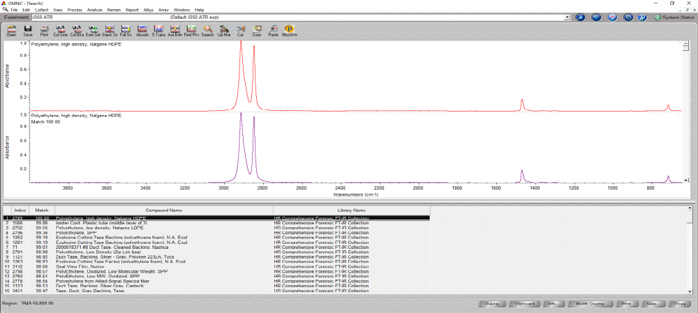
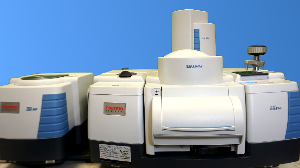
FTIR System Nicolet iS50 FT-IR analysis spectrometer: Our Nicolet iS50 is the center piece of our laboratory, as our primary analytical method. This is a cutting edge, top of the line system used in the identification of polymers, rubbers, pharmaceuticals, forensics or any other materials. We are able to identify foreign materials with ease, as…
LSB Products
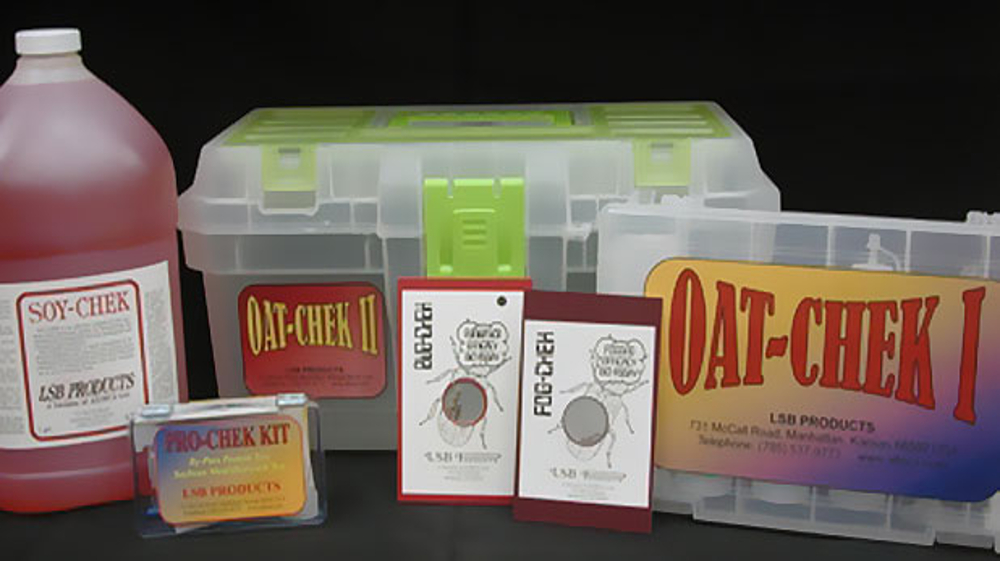
LSB Products manufactures rapid Quality Assurance test kits for use in food, feed and various processing and manufacturing operations. We have test kits to determine the cook quality of soybean, rice, oats and other products for use as foods and feeds. Our QC kits determine urease activity, by-pass protein, and hydrolytic and oxidative rancidity. BUG–CHEK…

Coming Soon…
Exciting new laboratory services are coming soon to Alteca. Stay tuned!